


A. Kebkalo, V. Hrianyla, A. Reiti, I. Yatsyk
Shupyk National Medical Academy, Kyiv
Introduction. Peritoneal dialysis is a convenient and effective method of intracorporeal detoxification for patients with chronic kidney disease [9]. The undeniable advantages of the method are the simplicity, the ability to have sessions of treatment at home, and its affordability. However, the method has a significant imperfection – the depletion of the transporting capacity of the peritoneum and its sclerosis. Infectious processes with an inert passing accelerate the functional exhaustion of the peritoneum significantly [3]. But even without infectious complications, the constant impact of substances used for dialysis leads to sclerosis and blockage of the peritoneal membrane’s transport abilities. It is known that there are parts of the peritoneum where the secretory and resorption are hundreds of times more active, for example, the recto-uterine pouch and the sub-diaphragmatic space, which play the biggest role in effective peritoneal dialysis. The data from the following literature describe cases of successful and prolonged course of peritoneal dialysis in patients with the isolation of a large area of the peritoneum because of the adhesive process [5, 6].
We suggested to apply laparoscopic isolation (saving the upper part of the peritoneum), using laparoscopic fixation of the greater omentum to the parietal peritoneum at the level of the umbilical ring. The main problem in this case is the formation of hermetic peritoneum parts without the connections with each other [2], which can be achieved by laparoscopic fixation of the greater omentum to the parietal peritoneum at the level of the umbilical ring.
The aim of this research. Our goal is to experimentally improve the method of fixation of the greater omentum to the parietal peritoneum to prolong the peritoneal dialysis procedure.
Materials and methods of the research. An experimental study was conducted on 60 Wistar rats. The requirements of bioethics, which are in line with the provisions of the European Convention for the Protection of Vertebrate Animals and the Law of Ukraine “On the Protection of Animals from Abuse,” were followed during the experiment. The main group of animals used for the research was formed by 30 rats, which had their peritoneums split into the upper and lower parts by a patented laparoscopic stapler. This device functions using bio-welding technologies (Fig. 1).
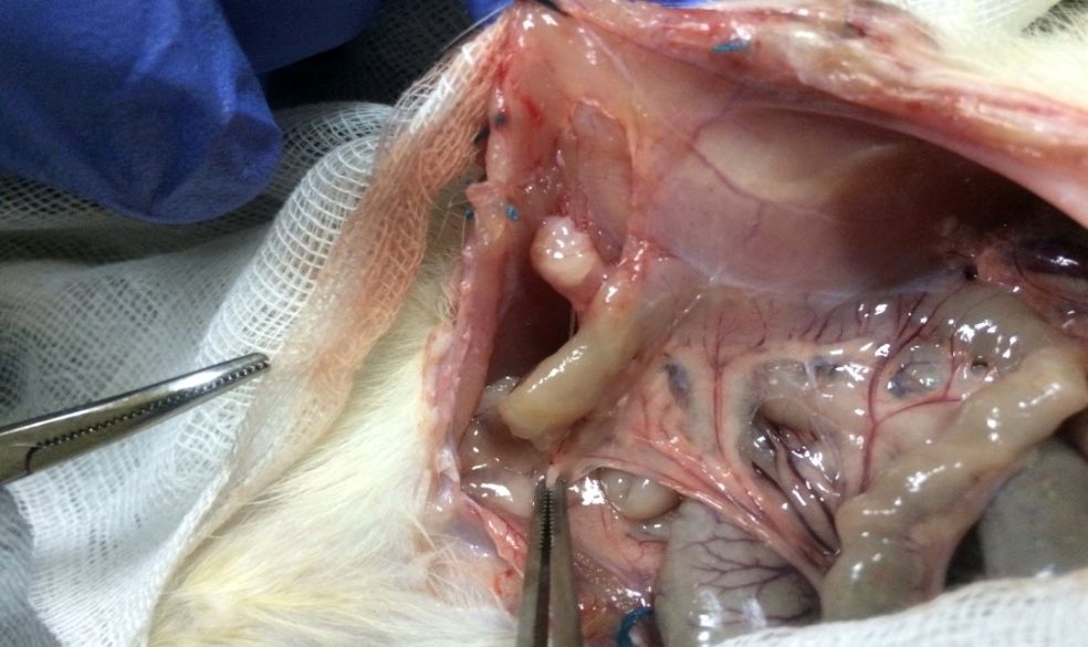
Fig. 1. Dividing of the peritoneum into parts using bio-welding technology.
30 rats from the controlled group had their peritoneums split into the upper and lower parts by suturing the greater omentum to the parietal peritoneum with the polyglycolide ligature the size of 4-0.
All the rats had two separate soft drainage put through the back into the upper and lower parts of the abdominal cavity [6]. In 14 days, 5.0 ml of 0.9 % saline, colored with brilliant blue, were injected through the “lower” dranaige. 3.0 ml of 0.9 % saline were injected through the “upper” one. There was no tightness, if after the aspiration the brilliant green appeared. The strength of the formed scar was evaluated too, both functionally and histologically.
To check the function of the scar the level of pressure that ruins the tightness of two parts of the peritoneum was tested. In order to do this, 5.0 ml of the colored solution was injected into the lower part. After that the aspiration from the upper part was done. The algorithm was repeated for several times.
The time needed for connection the greater omentum to the peritoneum, their histological abilities to recover on the 1st, 3rd, 7th and 14th days of the experiment were also compared.
The results of the study and the discussion of them. The average duration of the connection of the greater omenrum to the peritoneum in this experiment was 19.0 ± 70.0 min in the main group, and 35.0 ± 14.0 min in the controlled one. The time needed to form the described connection between the structures in the controlled group was longer, because of the frequent eruption of the delicate tissues by the ligature, the necessity of additional nodal sutures in places of insufficient tightness, whereas in the main group nothing like that happened due to using the special device. If the made connection was not strong enough, the tissue of the great omentum was moved 0.5 cm above or below the used area (Figs. 2, 3).

Fig. 2. Dividing of the peritoneum into parts using bio-welding technology.

Fig. 3. Dividing of the peritoneum into parts using suturing.
There was no difference in the amounts of leukocytes in the blood of both groups of the rats at day 1 and 3. However, on day 7 more leukocytes were found in the blood of the rats from the main group, on day 14 the levels of white blood cells were almost the same with a little bit bigger amount in the blood of rats from the main group (Fig. 4).

Fig. 4. The number of leukocytes in the blood of rats, g/l.
Histological characteristics: the necrotic scab with the active leukocyte infiltration was clearly visualized in the tissues taken from the rats of the main group; the rats of the controlled group had minor inflammatory processes and leukocyte infiltration histologically found in their tissues. (Fig. 5).

Fig. 5. Histological characteristics of the leukocyte infiltration.
On the 3rd day after surgery in the rats of the main group, there were found active phagocytosis of the scab because of burning tissies, many leukocytes and erythrocytes, signs of proliferation with fibroblasts at the exact place of the fixation, whereas in the rats from the compared group, leukocytes, moderate number of macrophages and single erythrocytes were explored (Fig. 6).

Fig. 6. Histological characteristics on the day 7 of the experiment.
On the 7th day in the main group active proliferative processes, disappearing of the burn scab, creating of infiltrating and proliferative area with an abundant vascularity, multiple fibroblasts were determined. At the same time, histological signs were unchanged and several blood vessels began to appear in the controlled group.

Fig. 7. Histological characteristics on the day 7 of the experiment.
On 14th day there were mature connective tissue elements along with young fibrocytes, elastic fibers and fibroblasts found in the main group.
In the controlled group, weakly expressed areas of creating of the connective tissue around the ligature were observed (Fig. 8).

Fig. 8. Histological characteristics on the day 14 of the experiment.
Thus, the strong connection of the greater omentum to the parietal peritoneum was created in the rats from the main group, whereas the connection between two structures in the rats from the controlled was kept only due to the ligature.
Comparison of tightness of the tissues of the rats from the main group showed a satisfactory result in 48 cases (96.0 %), in the rats from the controlled group – in 32 (64.0 %) (Fig. 9).

Fig. 9. The tightness of the peritoneum parts in the reats, %.
Comparison of the functional abilities of the connections showed that the starting point of ruining of the tightness in the main group started from 7.0 ml, and 100.0 % ruinig of the tightness occurred when using 11.5 ml. In the controlled group, the ruining of the tightness started from 6.0 ml and 100.0 % occurred after using 8.5 ml.
Conclusions. Using the bio-welding device improves the technique of the isolation of the abdominal cavity for prolonged peritoneal dialysis and provides for the creating of the reliable connection between the greater omentum and the parietal peritoneum, which is 74.5 % stronger than the one, created with ligature.
References
- Bazaev NA, Dorofeeva NI, Grinvald’VM, Putrya BM, Zhilo NA. Animal Trials of Wearable Apparatus for Peritoneal Dialysis. Journal Biomedical Radioelectronics. 2018;(6):12-14. (Russian).
- Bereshchenko VV, Voruschenko AV. Surgical Interventions in Patients Undergoing Peritoneal Dialysis. V: Materials of international scientific conference devoted to the 83rd anniversary of the Kursk state. med. u-ty “University Science: Look into the Future”. Kursk, 2018 Feb 02. Kursk, 2018:328-330. (Russian).
- Vatazin AV, Zulkarnaev AB, Rusanova EV, Budnikova NE. Bacterial and fungal pathogens in the transplantation and dialysis center. analysis for eighteen years (1998–2015). Russian Journal of Transplantology and Artificial Organs. 2016;18(2):56-64). (Russian). https://doi.org/10.15825/1995-1191-2016-2-56-64
- Helfand BR, Protsenko DN, Podachyn PV et al. Syndrome of Intraabdominal Hypertension: method. recommendations. Savelyeva VS, editor. Novosibirsk: Siberian Success; Partners of Siberia, 2008. 32 p.). (Russian).
- Zhura AV, Tretyak SI, Hryischanovich VYa, Makarevich ZhA. An experimental model of peritoneal adhesions. Experimental Surgery. 2017;25(4):333-339. (Russian). https://doi.org/10.18484/2305-0047.2017.4.333
- Poroyskiy SV, Poroyskaya AV, Bulycheva OS. Morphometric characteristics of the parietal and visceral peritoneum in the dynamics after various sizes surgical operation trauma application. Journal of VolgSMU. 2014;51(3):102-107. (Russian).
- Savytskyi IV, Tshipoviaz SV, Belash ОV, Vastyanov RS, Znamerovskyi SG, Lenik RG et al. Investigation of hematological indices in experimental peritonitis. Klinichna Khirurgiia. 2018;85(6):63-66. (Russian). https://doi.org/10.26779/2522-1396.2018.06.63
- Bradley SE, Bradley GP. The effect of increased intra-abdominal pressure on renal function in man. J Clin Invest. 1947;26:1010- https://doi.org/10.1172/JCI101867
- Mehrotra R, Devuyst O, Davies SJ, Johnson DW. The Current State of Peritoneal Dialysis. J Am Soc Nephrol. 2016; 27(11):3238-3252. https://doi.org/10.1681/ASN.2016010112